Rocks, Minerals, & Gems
Cathodoluminescence (CL) microscopy reveals the real structure of solids based on a crystal's intrinsic properties and trace-element chemistry. Although CL is useful for identifying minerals (and their distribution), researchers more frequently employ this technique to reveal the typomorphic properties and features of minerals that characterize the conditions of formation and alteration to reconstruct geological processes.
The table below shows various minerals that may be examined using CL. Note that iron minerals and iron-rich phases are generally non-luminescent.
| Class | Examples |
|---|---|
| Elements | Diamond |
| Sulfides | Sphalerite |
| Oxides | Corundum, cassiterite, periclase |
| Halides | Fluorite, halite |
| Sulfates | Anhydrite, alunite |
| Phosphates | Apatite |
| Carbonates | Calcite, aragonite, dolomite, magnesite |
| Silicates | Feldspar, quartz, zircon, kaolinite |
Experimental briefs and application notes

Webinar
Cathodoluminescence Explained. Episode 3: Analysis Modes for Geoscience Applications
Meteorites
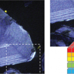
Meteorites can reveal fragments of the history of the solar system's evolution. With many long-term asteroidal sampling missions, petrographic investigations of meteorite specimens will assist in studying asteroid samples. These rocky carbonaceous aggregates include snapshots of the early solar system and can contain calcium aluminum inclusions (CAIs) and corundum grains, which can be among the first solids to condense from the solar nebula. Understanding their complex formation history and subsequent thermodynamic events is critical in resolving early planetary stages. The growth patterns, grain boundaries, and subtle zonation from crystallization can indicate formation from nebular gasses. Cathodoluminescence investigation of a meteoritic specimen can reveal and map the presence of trace elements and grain boundaries and assist with mineralogical identification.
 |
Sedimentary Rocks
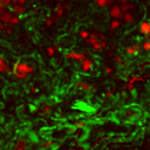
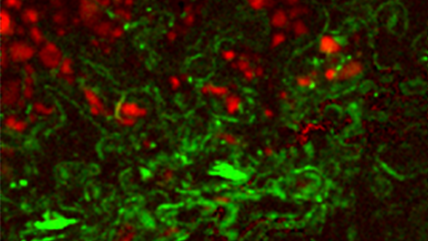
Sedimentologists use cathodoluminescence (CL) in clastic and carbonate petrography to reveal textural information that is not apparent with other (imaging) techniques. By studying grain provenance, sediment, and cement growth fabrics, CL studies provide insight to understand the diagenetic history better.
Cementation and diagenesis
Depositional grains and cement in carbonate and quartz minerals form under very different conditions, resulting in clear differences in their trace element chemistry. CL maps reveal the details of the growth fabric zonation, dissolution, and micro-fracturing, including successive cement generations.
In sandstone, detrital (of various origins) and authigenic quartz overgrowths enable quantification of cement abundance and determination of porosity-loss processes.
Provenance
CL is a valuable technique for monitoring the provenance of feldspars, mudrocks, and quartz (plutonic, volcanic, hydrothermal, and metamorphic). Specifically, maps can detect changes in crystal texture that correlate with impurity distributions and concentrations approaching parts per million levels.
References
Götze, J. M.; Plötze, M.; HaberMann, D., Mineral Petrol 71 (2001) p225 – 250
Scholonel, C.; Augustsson, C., Sedimentary Geology 336, 1 May 2016, p36 – 45
Color cathodoluminescence images captured using a ChromaCL2™ detector and provided courtesy of Prof. J Schieber, Indiana University Shale Research Lab.
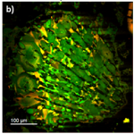
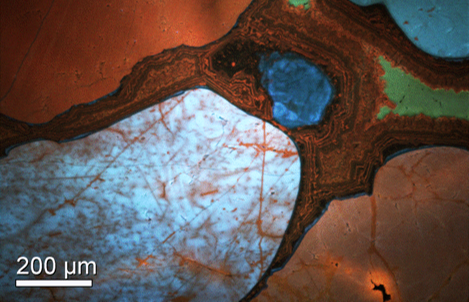
Carbonate Rocks
Petrography of carbonate rocks
Cathodoluminescence (CL) discriminates diagenetic relationships in carbonates, especially dolomite. CL can reveal complex growth histories of calcite and dolomite crystals and replacement relationships. Phosphorescence of the dominant (orange-red) luminescence of the manganese cation prevents the use of certain acquisition modes. However, wavelength-filtered or spectrum imaging approaches can be useful for visualizing the texture apparent at other wavelengths.
Economically Important Minerals and Gems
Reservoir rock
The oil and gas industry uses cathodoluminescence (CL) to characterize reservoir rocks.
Rare earth elements (REEs)
The last 20 years have seen an explosion in demand for REEs due to their use in rechargeable batteries, magnets, military-grade metal alloys, and many optoelectronic devices. Minable concentrations of REE-bearing minerals, such as apatite and monazite, are uncommon, and market availability is sometimes restricted.
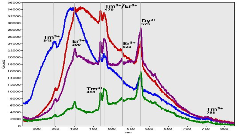
CL can reveal the presence of and identify REE within a mineral. Based on each REE's distinct emission spectrum, their distribution(s) may be mapped even at concentrations lower than ten parts per million.
Porphyry
Porphyry deposits are one of the world's most important copper, gold, and molybdenum repositories. High-resolution, scanning electron microscope CL mapping allows the detailed interpretation of the crystallization history within porphyry copper ore deposits to be understood.
Gems
Many gemstones, including diamonds and corundum (sapphire or ruby), exhibit strong CL and are widely studied. The inhomogeneous internal structure can reflect changes during growth and post-crystallization. This allows, for example, natural and synthetic diamonds to be distinguished based on their spectrum and a detailed understanding of the various colors they exhibit.
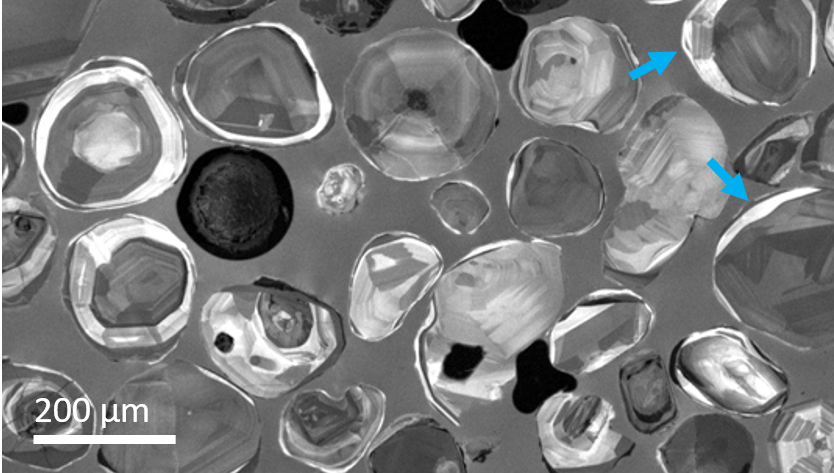
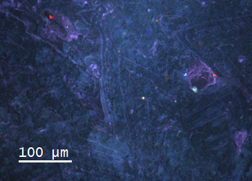
Quartz
One of the most important applications of cathodoluminescence (CL) in geosciences is the possibility of revealing internal structures, growth zoning, and lattice defects in quartz crystals that are not discernible by other analytical techniques. During quartz crystallization, variations of the physicochemical conditions result in crystal zoning. Revealing these features using CL enables geoscientists to reconstruct the specific growth conditions or to reveal multiphase processes. The zoning in quartz can relate to variations in trace-element uptake or intrinsic defects caused by the effects of growth conditions.

Provenance
The dependence of the CL spectrum (color) of quartz from different origins permits the recognition of specific formation conditions (e.g., temperature and pressure). Qualitative studies encompassing a wide array of quartz forms have enabled a general provenance classification to be built based on the CL color, with guidelines in place for the classification as volcanic, plutonic, hydrothermal, and metamorphic quartz fabrics. However, in combination with other analytical methods such as trace-element analysis, isotope, or inclusion studies, it is often beneficial.
The concentration of titanium in quartz is widely demonstrated as a valuable tool for determining the temperature and pressure of quartz crystallization (the so-called TitaniQ method). Qualitative studies show that blue wavelengths could be associated with titanium concentration. However, more recently, it has been noted that spectrum imaging (hyperspectral imaging) approaches are necessary for accurate quantitative analysis.
Diagenesis
CL maps are useful for reconstructing diagenetic processes in reservoir quartz since authigenic and detrital quartz can be distinguished, as well as secondary overgrowths, ghosts of the original fabric, and cementation sequences. Such observations are important for discerning various geological and mineralogical aspects, particularly in oil exploration.
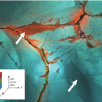
Metamorphism
CL is useful for unraveling deformation features (e.g., microfractures) or fluid migration pathways in crystalline and sedimentary rocks. For instance, it has been demonstrated that CL is an excellent tool for distinguishing brittle or ductile deformation processes during sandstone diagenesis. On the other hand, CL can reveal fluid migration due to hydrothermal alteration or pro- and retrograde metamorphic conditions often not recognized by conventional polarizing microscopy.
Zircons
Geochronology
Zircons are frequently used as geological clocks because they include small amounts of radioactive uranium isotopes and their decay products are incorporated into the crystal structure during crystallization. In old inherited zircon cores, younger igneous zircons, or ones that have undergone more than one crystallization event, the U/Pb isotope ratio in each crystallization period will differ based on the time elapsed (uranium is immobile in the zircon crystal). This polyphase structure is often not recognizable by conventional imaging methods, such as light microscopy, but is necessary for accurate dating results. However, zircons can be selected for dating purposes using cathodoluminescence (CL) in the scanning electron microscope. An unfiltered CL image is useful to identify zircon phases of different ages that appear as bright regions (e.g., the presence of late rims) at the grain perimeter. Moreover, conclusions on the origin and development of the inherited phases can be drawn by comparison with detrital zircons.