WHAT IS CL
  • Introduction
    • What is CL?
    • Why use CL?
    • Modes of Operation
      • Unfiltered
      • Wavelength-Resolved
      • Wavelength-Filtered
      • Angular-Resolved (ARCL)
      • Polarization
      • Time-Resolved
      • Wavelength- and Angle-Resolved (WARCL)
  • Applications
    • Rocks, Minerals, & Gems
      • Meteorites
      • Sedimentary Rocks
      • Carbonate Rocks
      • Economically Important
      • Quartz
      • Zircons
    • Semiconductors
      • Process Development
      • Nanostructure Characterization
      • Device Characterization
        • LEDs
        • MicroLED Displays
        • Laser Diodes
        • Photovoltaics
    • Nanophotonics
      • Plasmonics
      • Metamaterials
      • Photonic Structures
    • Ceramics, Oxides, & Nitrides
      • Phosphors
      • Oxides & Nitrides
      • Dielectric Materials
      • Engineering Ceramics
    • Radiations
    • Organic Molecules
      • Biological Specimens
      • Pharmaceuticals
      • Polymers
  • How To
    • Getting Ready
      • Sample Preparation
      • Microscope Setup
      • Alignment
        • For Monarc
        • For MonoCL and ChromaCL
        • For Vulcan
      • Technique Selection
    • Spatial Mapping
      • Alignment for Mapping
      • Collecting Maps
      • Optimizing Maps
    • Spectroscopy
      • Alignment for Spectroscopy
      • Collecting Spectra
      • Optimizing Spectra
    • Emission Patterns
      • Alignment for Emission Patterns
      • Collecting Emission Patterns
      • Optimizing Emission Patterns
    • Spectrum Imaging
      • Wavelength-Filtered SI
      • Wavelength/Angle-Resolved SI
        • SEM/STEM SI Setup
        • Survey Image
        • Select and Survey ROI
        • Flow Control
        • Visual Feedback
        • Adjustments
      • Advanced SI
    • Analyze
      • Origins of Contrast
      • Luminescence Spectra
      • Least-Square Fitting
        • Single Spectrum
        • Spectrum Image
        • NLLS Models
        • NLLS Setup Palette
      • Tips & Tricks
  • Resources
  • Events
  • Glossary
  • FAQ
  • Contact
  • Getting Ready
    • Sample Preparation
    • Microscope Setup
    • Alignment
      • For Monarc
      • For MonoCL and ChromaCL
      • For Vulcan
    • Technique Selection
  • Spatial Mapping
    • Alignment for Mapping
    • Collecting Maps
    • Optimizing Maps
  • Spectroscopy
    • Alignment for Spectroscopy
    • Collecting Spectra
    • Optimizing Spectra
  • Emission Patterns
    • Alignment for Emission Patterns
    • Collecting Emission Patterns
    • Optimizing Emission Patterns
  • Spectrum Imaging
    • Wavelength-Filtered SI
    • Wavelength/Angle-Resolved SI
      • SEM/STEM SI Setup
      • Survey Image
      • Select and Survey ROI
      • Flow Control
      • Visual Feedback
      • Adjustments
    • Advanced SI
  • Analyze
    • Origins of Contrast
    • Luminescence Spectra
    • Least-Square Fitting
      • Single Spectrum
      • Spectrum Image
      • NLLS Models
      • NLLS Setup Palette
    • Tips & Tricks
Back to Top

How to

Getting Ready

Getting Ready

Understand the basic requirements and setup necessary to acquire data with cathodoluminescence microscopy.

  • Sample Preparation
  • Microscope Setup
  • Alignment
  • Technique Selection

Spatial Mapping

See the recommended workflow to align the sample, CL system, and microscope that enable high-quality data collection in the shortest time.

  • Alignment for Mapping
  • Collecting Maps
  • Optimizing Maps

Spectroscopy

Follow step-by-step instructions to acquire cathodoluminescence spectral data to analyze your sample.

  • Alignment for Spectroscopy
  • Collecting Spectra
  • Optimizing Spectra

Emission Patterns

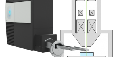
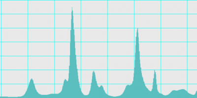
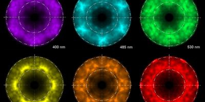
Understand angle-resolved (AR) and wavelength- and angle-resolved (WAR) CL best practices to acquire the highest quality emission patterns from materials and devices.

  • Alignment for Emission Patterns
  • Collecting Emission Patterns
  • Optimizing Emission Patterns

Spectrum Imaging

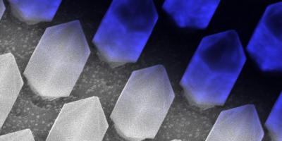
Explore how to collect combined spatial and spectroscopic data so you can utilize the power of CL spectral analysis with the spatial resolution of an electron microscope.

  • Wavelength-Filtered SI
  • Wavelength/Angle-Resolved SI
  • Advanced SI

Analyze

Learn how to use Gatan Microscopy Suite® (GMS) to process and analyze CL spectra and spectrum images.

  • Origins of Contrast
  • Luminescence Spectra
  • Least-Square Fitting

Want to enhance your CL results?

Access application experts who are ready to help solve your CL issues!

Contact Us
Gatan logo
  • Gatan Corporate
  • Privacy Policy
  • Events
  • Glossary
  • FAQ
  • Contact
Join the community
Back to Top