Spectrum image display
During spectrum image (SI) acquisition, each SI is shown in its own image display. The spectrum image fills as the spectral data is acquired and placed into position in the SI. You can use the 3D visualization tools to explore the spectral data while the acquisition is running (e.g., the Slice tool). Since a processing overhead may introduce artifacts into your data acquisition, we advise doing this after acquisition or while paused.
Live spectra
Live spectral feedback during acquisition can be enabled or disabled with the Live button in the STEM-SI palette. You can manipulate the live spectra using standard line plot display visualization tools.

Beam position cursor
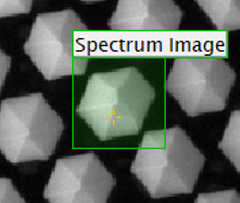
During acquisition, an orange beam cursor marks the beam position on the survey image. This indicates the progress of the acquisition, and its position may vary slightly from that of the beam. For particularly fast acquisitions, the cursor is displayed as a line or even completely disabled.
Pixels per second
![]() The actual SI acquisition rate is posted to the DigitalMicrograph® software status area at the bottom of the application. It displays in units of pixels per second. This information can be useful when configuring a spectrometer for optimal readout speed.
The actual SI acquisition rate is posted to the DigitalMicrograph® software status area at the bottom of the application. It displays in units of pixels per second. This information can be useful when configuring a spectrometer for optimal readout speed.
Time remaining
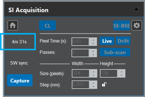
The remaining acquisition time is displayed in real-time in the STEM-SI palette above the Capture button. This time is based on the actual acquisition rate, excluding any pauses.