Acquire a spectrum
 There are two methods to collect a cathodoluminescence (CL) spectrum: wavelength-resolved and wavelength-filtered spectroscopy. The appropriate method depends on the type of detector you use. Please refer to the modes of operation section for more information.
Within DigitaMicrograph® software, go to the Spectroscopy technique to capture CL spectra:
 Position the beam on the sample region of interest using the Scan palette.
- Spot – Sets the beam at a particular location on (or to avoid damage, off of) the specimen.
- Focus – Scans a sub-region of the field of view.
Ensure that the refresh rate of the scan is shorter than the exposure time of the spectrum.
- Capture a spectrum.
Wavelength-resolved
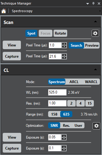
Use the CL palette to capture a wavelength-resolved spectrum. Simply press View or Capture to collect a CL spectrum.
- View – The spectrum continually updates per the exposure time selected
- Capture – Records a single CL spectrum, then opens the data and includes the SEM's electron beam location in a new workspace
The user interface allows you to control the capture of spectral information:
- WL (nm) – Designates the central wavelength of a CL spectrum
- Res. (nm) – Defines the instrument's spectral resolution (wavelength)
- Range (nm) – Select the appropriate button to choose the (wavelength) field of view in a CL spectrum; this selects a diffraction grating of different dispersion
- Optimization – Determines the read-out configuration of the detector
- SNR – High signal-to-noise
- Res. – High resolution
- User – User-defined binning and dark correction
The Spectrum Imaging section includes additional information regarding how to collect spatial and spectral information using the wavelength-resolved spectrum imaging mode.
 Wavelength-filtered
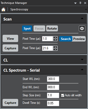
Use the CL Spectrum – Serial palette to capture a wavelength-filtered spectrum. Similar to above, simply press Capture to collect a CL spectrum.
- Start WL (nm) – Designates the short wavelength
- End WL (nm) – Defines the long wavelength
- Step Size (nm) – Select the wavelength interval to take each spectrum
When you select the Auto slit width checkbox, the system automatically sets the entrance and exit slit widths to maximize spectral resolution and throughput for the user-selected sampling step. - Dwell Time (s) – Time spent at each wavelength
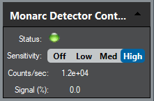
For systems that use a computer-controlled detector, you must select the correct detector and an appropriate sensitivity level from the Monarc/Vulcan Detector Control palette.
 The Wavelength-Filtered SI section includes additional information regarding how to collect spatial and spectral information using the wavelength-filtered spectrum imaging mode.