Showing 1-10 of 15
  • Media image

    Local surface plasmon resonance mode (LSPR) mapping of gold pyramids using cathodoluminescence. (a) Composite image of secondary electron image (gray) and, 650 nm (green) and 750 nm (red) monochromatic cathodoluminescence images.

  • Media image
    Data courtesy of Dr. U. Jahn, Paul-Drude-Institut für Festkörperelektronik.

    (a) Secondary electron image of a 200 nm diameter GaAs nanowire; (b) Cathodoluminescence spectrum-image displaying energy plane 1.46 ± 0.02 eV; (c) Individual cathodoluminescence spectrum extracted from spectrum-image with single peak at 1.45 eV; (d) Map of central energy of luminescence; change

  • Media image
    Data courtesy of Dr. E. Vicenzi, Smithsonian Institute

    (a) Composite cathodoluminescence image of polycrystalline diamond created using red, green, and blue spectrally filtered cathodoluminescence images. Radioactive fluid ingress along grain boundaries leads to radiation halo effects being observed (yellow).

  • Media image
    Data courtesy of Prof. Nicholas Leventis, Chemistry Department, Missouri University of Science and Technology

    Cathodoluminescence image of paint pigment sample prepared with Ilion II and imaged with MonoCL4™ system.

  • Media image
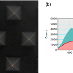
    Data courtesy of Dr. R. Williams, Ohio State University

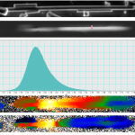
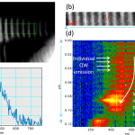
    Cathodoluminescence analysis of a GaN/AlN nanowire in STEM (a) Dark field STEM image, (b) cathodoluminescence spectrum-image acquired from the region indicated by the green rectangle in (a), (c) A cathodoluminescence spectrum extracted from the spectrum-image, and (d) a projection of the cathodol

  • Media image

    Panchromatic cathodoluminescence image of bulk GaN sample. Dark spots (some indicated by blue arrows) correspond to threading dislocations. A dislocation density of 5 x 105 cm-2 was measured. Acquisition time = 30 s.

  • Media image
    Data courtesy of G Nichols, Pfizer UK

    Distribution of drug within a pharmaceutical pill. Composite color image showing the spatial distribution of the micronized drug, salmeterol xinafoate.

  • Media image
    Data courtesy of Dr. M. Bosman, A*STAR IMRE, Singapore

    Cathodoluminescence investigation of gold prisms in the STEM: (a) and (b) are bright field TEM images and (c) and (d) are the cathodoluminescence intensity maps recorded in STEM mode from particles (a) and (b) respectively.

  • Media image
    Images (a), (b) and (d) –(f) courtesy of Dr. J. Schieber, Indiana University.

    Color cathodoluminescence reveals provenance of populations of quartz grains (a) Cleveland shale (Devonian); low grade metamorphic acadian orogeny; (b) Barnett shale (Miss.) Texas; low grade metamorphic ouchita orogeny; (c) Unspecified; (d) Santa Barbara basin shale (modern); plutonic (Sierra Nev

  • Media image
    Image courtesy of Dr. J. Schieber, Indiana University.

    Quartz grains from granite and metamorphic source with low temperature quartz cement (bluish). There is a later (hotter) overprint (reddish) along grain boundaries in the overgrowth.